Case 13 Answer: DVA (developmental venous anomaly)Yes, I was misleading with the history but unfortunately this was the real one. I don't...
Case 13 Answer: DVA (developmental venous anomaly)
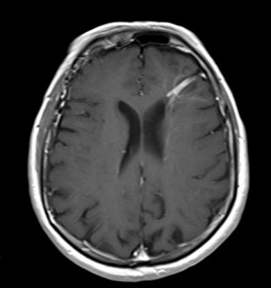
Yes, I was misleading with the history but unfortunately this was the real one. I don’t fault anyone for thinking it could be blood given what I told you, but is hemorrhage usually linear? and that deep in the white matter?
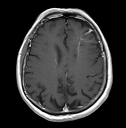
Classically DVAs are in the frontoparietal region and are best seen on CT/MRI but their large draining vein with runs centripetally towards the ventricle, often with some calcifications. It should stay unchanged on the follow up and if contrasted would enhance well. MRI shows the same thing, just better.
A few people thought of DAI but remember those should be more punctate and numerous, scattered around the grey-white junction. But again, always err on the side of caution till that f/u CT or MRI shows stability.
reference: https://radiopaedia.org/articles/developmental-venous-anomaly