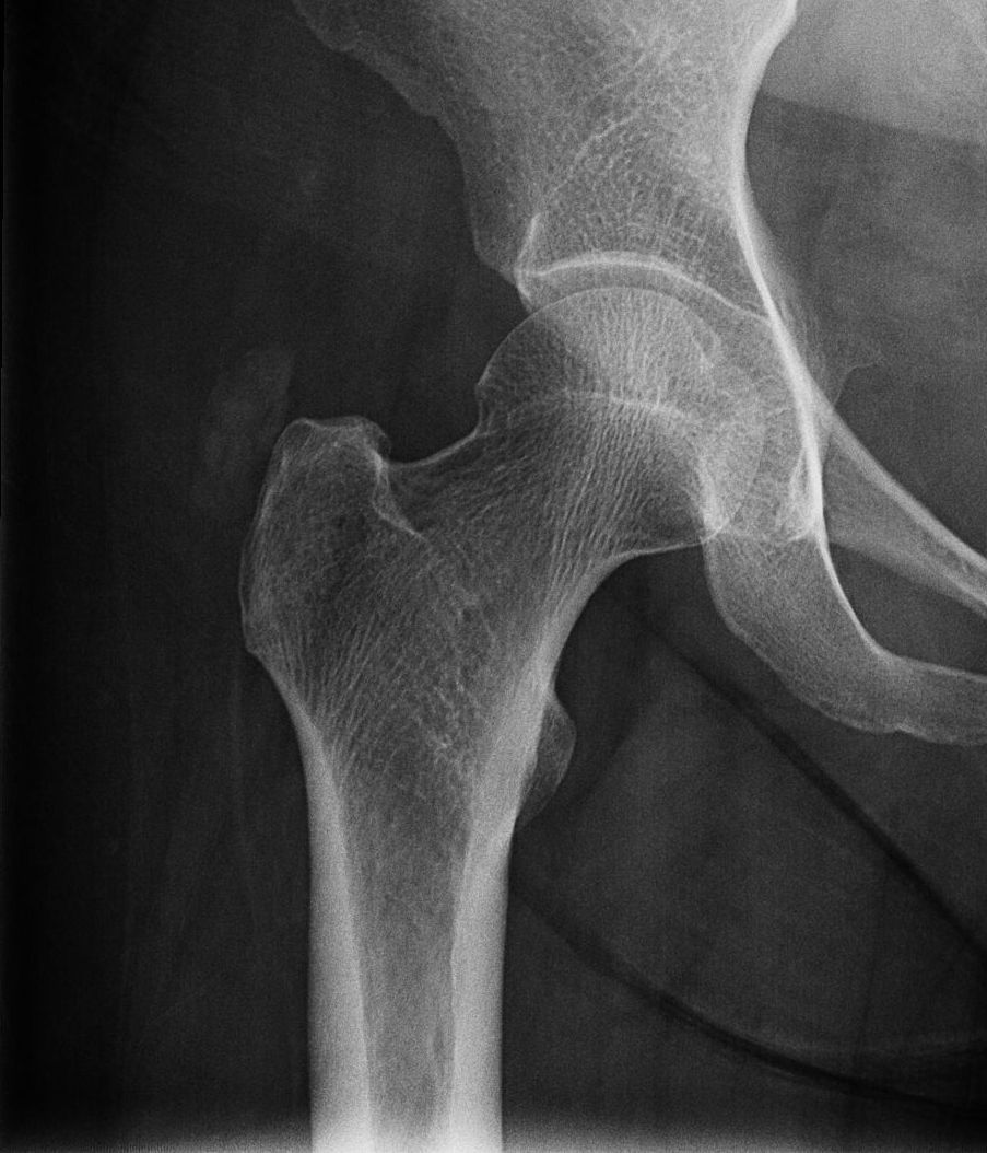
Case 24 AnswerHere's a more coned down view. Did you see the pathology?There is amorphous calcification over the greater trochanter. Where...
Case 24 Answer
Here’s a more coned down view. Did you see the pathology?
There is amorphous calcification over the greater trochanter. Where is it? Dig out your MSK anatomy. Generally there are two possibilities when you see such calcifications about a joint, a) within the tendons or b) within a bursa. Sometimes it can be difficult to tell on plain films, at which point an MRI would show it much better.
Answer: calcific bursitis of the trochanteric bursa versus calcific tendonitis of the gluteus medius tendon (favor bursitis)