

Season 9 Case 8
Hx: 30yo female w abdominal pain
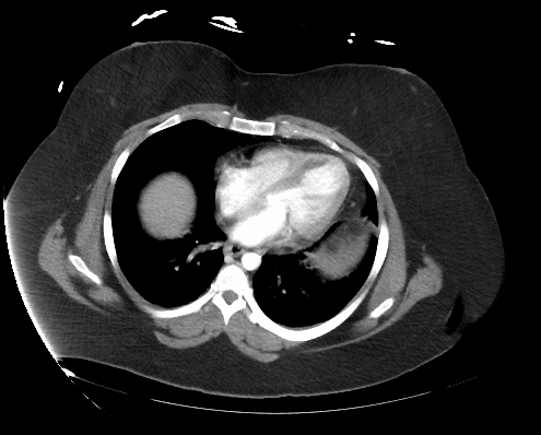
Answer: Hepatic Adenoma with hemorrhage
Multiple hyperenhancing hepatic masses in a young female. Largest in left lobe within internal hyperdensity (blood) and capsular irregularity consistent with extracapsular hemorrhage (hepatic rupture).


Hepatic Adenomas:
-Benign
-m/c hepatic tumor in young females classically on OCPs
-stimulated by estrogen (OCPs, obesity, anabolic steroid use)
-typically asymptomatic
-prone to hemorrhage which can even yield exsanguination!
Hepatic Adenomas Imaging:
Classic:
-solitary (>10 = adenomatosis)
-large (5-15cm) at dx
-subcapsular, R hepatic lobe
-well defined -Ca++ uncommon (old bleed)
-US: varies
-CT: isodense to liver, early enhancement but iso-enhancing by portal venous phase. Hyperdense in a fatty liver or when containing hemorrhage
MR: T1 variable, T2 mild increased, +fat ->phase drop out
-contrast: early arterial enhancement then iso on delay (like CT)
-hemorrhage can distort this
Nucs: photopenic defect on Tc-99 sulfur colloid ( increased number & function of Kupffer cells)
